Software Available
Epicardial Fat Segmentation Tool
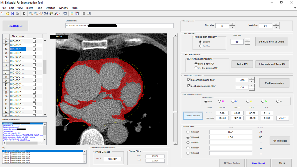
The Epicardial Fat Segmentation Tool supports the radiologists in the procedure of Epicardial Fat Volume (EFV) segmentation and quantification. By means of a user-friendly Graphical User Interface (GUI), the Tool takes as input a cardiac CT series and – with few simple steps and with operator intervention only in the initial phase – yields the EFV and automatically performs the quantification of the fat quartiles and their distribution around the heart. The whole processing pipeline for the EFV segmentation and quantification can be divided into 6 steps:
- ROI Selection
- ROI Interpolation
- ROI Refinement
- Epicardial Adipose Tissue Segmentation
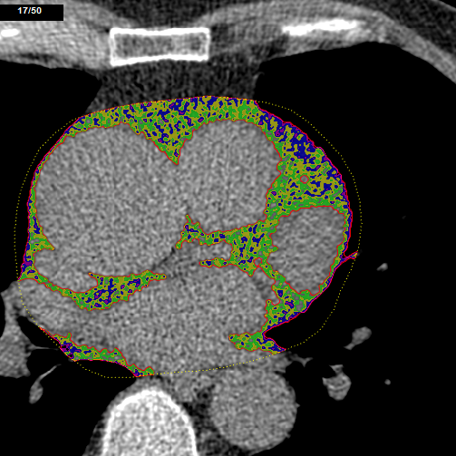
- Fat Volume and Quartiles Computation
- Fat Thickness Measurement.
Software Citations: 41
Please, if you are using the Epicardial Fat Segmentation Tool, please cite the following papers:
A semi-automatic approach for epicardial adipose tissue segmentation and quantification on cardiac CT scans
CT radiomic features and clinical biomarkers for predicting coronary artery disease
Epicardial and thoracic subcutaneous fat texture analysis in patients undergoing cardiac CT.
Militello, C., Rundo, L., Toia, P., Conti, V., Russo, G., Filorizzo, C., Maffei, E., Cademartiri, F., La Grutta, L., Midiri, M. and Vitabile, S.
Computers in biology and medicine, Elsevier. DOI
Militello, C., Prinzi, F., Sollami, G., Rundo, L., La Grutta, L. and Vitabile, S.
Cognitive Computation, Springer. DOI
Agnese, M., Toia, P., Sollami, G., Militello, C., Rundo, L., Vitabile, S., Maffei, E., Agnello, F., Gagliardo, C., Grassedonio, E. and Galia, M.
Heliyon, Elsevier. DOI
For more details contact: Prof. Eng. Salvatore Vitabile or Eng. Carmelo Militello

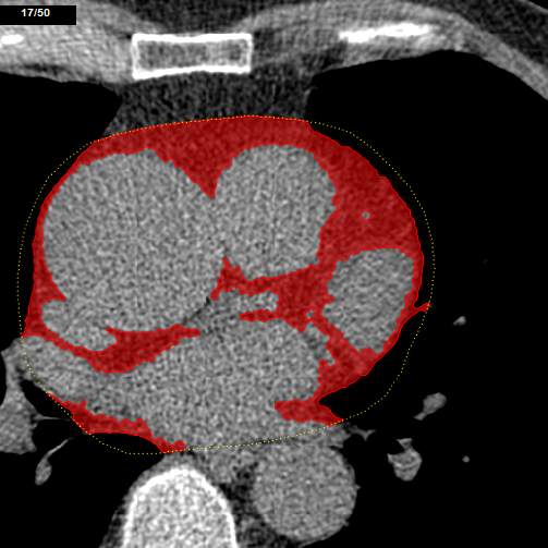
The GUI of the implemented tool for the EFV segmentation and quantification. On the left side, the list of all the slices belonging to the loaded CT series is displayed. In the central area, the selected image is displayed by over-imposing (transparency obtained by using alpha blending) the result of the performed segmented EFV. On the right side, the controls necessary to perform the various processing steps are shown.

